
The HIFα-Stabilizing Drug Roxadustat Increases the Number of Renal Epo-Producing Sca-1+ Cells
Aline Jatho , Anke Zieseniss , Katja Brechtel-Curth , Jia Guo , Kai Oliver Böker , Gabriela Salinas , Roland H Wenger, Dörthe M Katschinski
Inhibition of the prolyl-4-hydroxylase domain (PHD) enzymes, leading to the stabilization of hypoxia-inducible factor (HIF) α as well as to the stimulation of erythropoietin (Epo) synthesis, is the functional mechanism of the new anti-anemia drug roxadustat. Little is known about the effects of roxadustat on the Epo-producing cell pool. To gain further insights into the function of PHD inhibitors, we characterized the abundance of mesenchymal stem cell (MSC)-like cells after roxadustat treatment of mice. The number of Sca-1+ mesenchymal cells following roxadustat treatment increased exclusively in the kidneys. Isolated Sca-1+ cells demonstrated typical features of MSC-like cells, including adherence to tissue culture plates, trilineage differentiation potential, and expression of MSC markers. Kidney-derived Sca-1+ MSC-like cells were cultured for up to 21 days. Within the first few days in culture, cells stabilized HIF-1α and HIF-2α and temporarily increased Epo production upon incubation in hypoxia. In summary, we have identified a Sca-1+ MSC-like cell population that is involved in renal Epo production and might contribute to the strong anti-anemic effect of the PHD inhibitor roxadustat.
Precisely Tuned Inhibition of HIF Prolyl Hydroxylases Is Key for Cardioprotection After Ischemia
Aline Jatho , Anke Zieseniss, Katja Brechtel-Curth, Atsushi Yamamoto, Mathew L Coleman, Ana M Vergel Leon, Daniel Biggs, Ben Davies, Chris W Pugh, Peter J Ratcliffe, Dörthe M Katschinski
PMID: 33626887
PMCID: PMC8048377
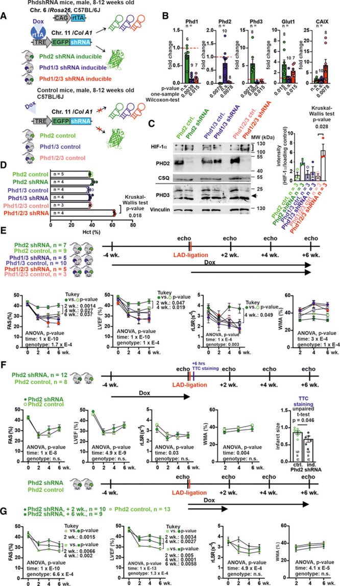
Acute myocardial infarction (AMI) is associated with a high incidence of heart failure and mortality. The HIF (hypoxia-inducible factor) system plays a central role in the adaptation to limited oxygen supply and may, in some circumstances, protect against ischemic damage. The protein stability of the HIFα subunits is regulated by 3 oxygen- and iron-dependent PHD (prolyl-4-hydroxylase-domain) enzymes, which are druggable targets.1 Roxadustat and daprodustat are first-in-class PHD inhibitors with regulatory approval in China and Japan for renal anemia treatment. They induce endogenous erythropoietin expression by inhibition of all 3 PHD enzymes. Repurposing PHD inhibitors for tissue protection is an attractive strategy, which depends on the possibility of achieving anti-ischemia effects without excessive erythrocytosis. This raises questions regarding PHD isoform-specific effects and timing of treatment. The issue has not been addressed before since the currently available PHD inhibitors lack isoform-specificity.
RhoA Ambivalently Controls Prominent Myofibroblast Characteritics by Involving Distinct Signaling Routes
Aline Jatho , Svenja Hartmann, Naim Kittana, Felicitas Mügge, Christina M Wuertz, Malte Tiburcy, Wolfram-Hubertus Zimmermann, Dörthe M Katschinski, Susanne Lutz
Affiliations Expand
PMID: 26448568
PMCID: PMC4598021
Abstract
Introduction: RhoA has been shown to be beneficial in cardiac disease models when overexpressed in cardiomyocytes, whereas its role in cardiac fibroblasts (CF) is still poorly understood. During cardiac remodeling CF undergo a transition towards a myofibroblast phenotype thereby showing an increased proliferation and migration rate. Both processes involve the remodeling of the cytoskeleton. Since RhoA is known to be a major regulator of the cytoskeleton, we analyzed its role in CF and its effect on myofibroblast characteristics in 2 D and 3D models.
Results: Downregulation of RhoA was shown to strongly affect the actin cytoskeleton. It decreased the myofibroblast marker α-sm-actin, but increased certain fibrosis-associated factors like TGF-β and collagens. Also, the detailed analysis of CTGF expression demonstrated that the outcome of RhoA signaling strongly depends on the involved stimulus. Furthermore, we show that proliferation of myofibroblasts rely on RhoA and tubulin acetylation. In assays accessing three different types of migration, we demonstrate that RhoA/ROCK/Dia1 are important for 2D migration and the repression of RhoA and Dia1 signaling accelerates 3D migration. Finally, we show that a downregulation of RhoA in CF impacts the viscoelastic and contractile properties of engineered tissues.
Conclusion: RhoA positively and negatively influences myofibroblast characteristics by differential signaling cascades and depending on environmental conditions. These include gene expression, migration and proliferation. Reduction of RhoA leads to an increased viscoelasticity and a decrease in contractile force in engineered cardiac tissue.